AI 생성 일러스트레이션
로그인
AI 생성 일러스트레이션
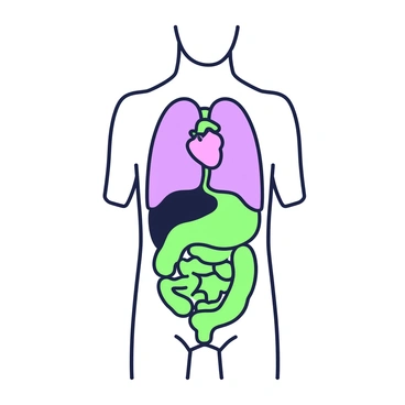
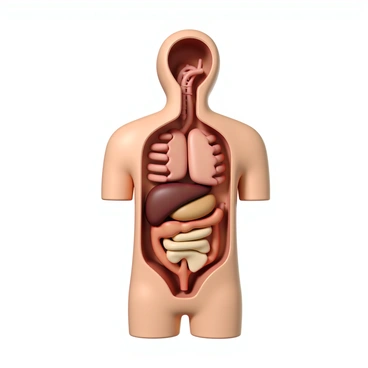
Body
Identify cavities following
Identify cavities following
커뮤니티에서 검색된 내용이 없습니다
나의 생성